contact lens case reports
BY PATRICK J. CAROLINE, FAAO, & MARK P. ANDRÉ, FAAO
Fitting a Semi-scleral Lens on a Highly Irregular Cornea
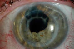
Patient JH is a 59-year-old male who was hit with a metallic foreign body that penetrated his left cornea, iris and lens in 1981. The wound was surgically repaired, and in 1983 surgeons removed the moved the traumatic cataract and replaced it with an anterior chamber IOL. Over the next 20 years the cornea began a slow but progressive course of decomposition, and he subsequently underwent a penetrating keratoplasty in March 2005. The transplant was healing uneventfully until the patient suffered a wound dehiscence that required additional surgical intervention. This series of events left JH with a clear but highly irregular and asymmetrical cornea (Figure 1).

Figure 1. Patient JH post keratoplasty and wound dehiscence.

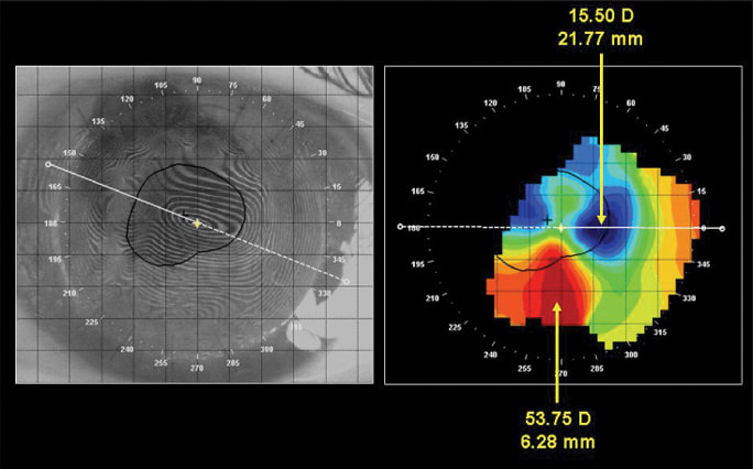
Figure 2. Left eye photokeratoscopy and corneal topography.
Drs. Beth Kinoshita and Matthew Lampa at the Pacific University Therapeutic Contact Lens Clinic examined JH in September 2006. At that visit the patient’s right eye was 20/20 with minimal refractive error of +0.50 -0.50 ×170 and the left eye was 20/800 uncorrected. Corneal mapping showed a normal right eye. The post-keratoplasty left eye demonstrated a high, irregular, with-the-rule corneal astigmatism with the steepest portion of the cornea inferiorly at 53.75D (6.28mm) and the flattest paracentrally at 15.50D (21.77mm) (Figure 2).
Finding the Right Lens
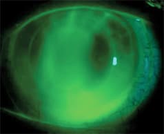
After trying a number of more traditional post-keratoplasty diagnostic lenses, Drs. Kinoshita and Lampa determined that a 15.4mm semi-scleral Jupiter Lens (Medlens Innovations) might be the best option. Because of the highly irregular, oblate cornea, they ordered a reverse geometry Jupiter Lens with the following parameters: 32.00D (9.38mm) base curve, +11.00D, 15.4mm diameter, 44.00D (7.67mm) mid-peripheral alignment curve and a reverse curve radius of 6.50mm.
JH reported diplopia upon lens application, but it diminished over the next two weeks as binocular vision reestablished. A single power adjustment brought the visual acuity to 20/50 (Figure 3). At JH’s last visit in March 2007, his visual acuity with the Jupiter contact lens was 20/40 and he enjoyed all day comfort and wearing time.

Figure 3. Fluorescein pattern of the reverse geometry Jupiter Lens.
This case illustrates the distinct advantages of semi-scleral lens designs that can help center and support the lens on the normal scleral curve while vaulting the highly astigmatic and asymmetrical corneal surface. CLS
| Patrick Caroline is an associate professor of optometry at Pacific University. He is also a consultant to Paragon Vision Sciences and SynergEyes, Inc. Mark André is an associate professor of optometry at Pacific University. He is also a consultant for Alcon Labs, CooperVision and SynergEyes, Inc. |


